Anatomy Appeal: The surgeon's roadmap
Project update from the illustrator - November 2017
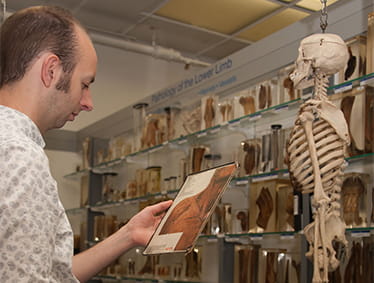
Drawing anatomical specimens is a process of revealing the workings of the body. I am not only drawing the form of the specimen, but also drawing the viewer’s eye through the chaos of tissue to show order and understanding of the organ or muscle. To achieve this I use the many qualities of pencil drawing: shading to reveal form and firmness, line to reveal distinction and strength. This is where traditional techniques of pencil drawing beat more modern computer based approaches to medical illustration: the image from a computer model, though multi-coloured to help the eye, is still little more mediated than a photograph itself.
My own interest in anatomy came through drawing. As a masters student at the Royal College of Art I discovered facial reconstruction, which I later studied at Dundee and through that found a deep interest in anatomy where I then went on to study an MSc in Human Anatomy. As Prosector at Brighton and Sussex Medical School, Sussex University I maintain an ongoing relationship with work on human anatomy, reaffirming an understanding which could not be gained from a living subject and which, in turn, re-informs my drawing.
by Lydia Carline
Help us create new anatomical illustrations for surgical training
With your generous support we have now raised £12,362 of our £15,000 Christmas target. Donate today to help us achieve our target.
 Thousands of medical trainees and professionals visit the Wellcome Museum of Anatomy and Pathology at the RCS every year. It's a unique, national teaching and study centre with a vast number of anatomical and pathological specimens collected over the past 200 years.
Part of the collection’s unique value comes from the wide array of examples of specific pathologies that are an important teaching aid in understanding diseases which present in clinic. However:
"The educational value of many of the specimens is significantly diminished because they lack an illustrated key to aid identification of specific structures. Labelled photographs are available for a selection of the potted specimens, but interpreting complex dissections, even with the aid of a photograph, can be difficult: a drawing conveys anatomical relationships with greater clarity and achieves an impression of three dimensionality more successfully than a photograph." - Professor Susan Standring, Professor of Anatomy and author of the latest edition of Gray’s Anatomy
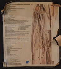 Specimens are currently supported by labelled photograph cards. These photographs were extracted from text books in the 1980s and mounted on cardboard. After 35 years of regular use these cards are in poor condition; most are torn and have large areas of damage and staining like the example on the right.
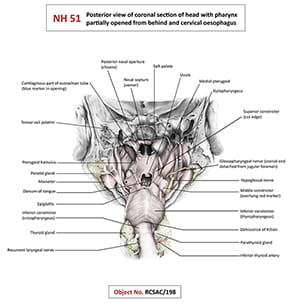
Replacing the current photographs is an option, but a series of detailed anatomical illustrations would provide a much better and more effective teaching aid.
Unlike photographic images, high-quality diagrammatical illustrations present a clear, easy-to-interpret image of complex anatomy. Once captured digitally, these illustrations would have an enduring lifespan and multiple uses in book illustration and for web-based educational material.
Support our appeal
 We'd like to commission 25 illustrations of the most frequently-used specimens in the Wellcome Museum of Anatomy and Pathology.
Each illustration will take approximately four days to complete (depending on the complexity of the specimen) at a cost of £600 per illustration. This includes the cost of labelling and consultation with teaching faculty. We'd be delighted to recognise financial support by including the name and a suitable acknowledgement on each illustration sponsored.
Thanks to the support of our friends and donors we have now commissioned 20 of these illustrations. Please donate to help us to reach our target of 25.
How to donate
Please donate online, or fill out and return a donation form to the address below:
RCS Anatomy Appeal donation form
Development Office
The Royal College of Surgeons of England
FREEPOST WC5162/2
London WC2A 3PE